

X-Ray Interpretation
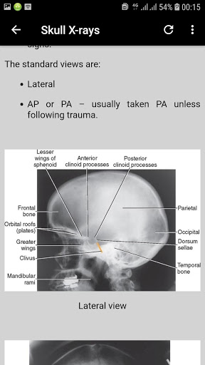
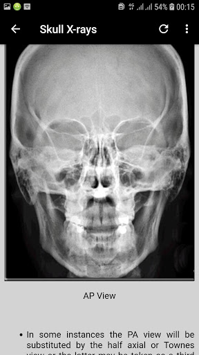
Interpretation of Skull, paranasal sinuses, orbit, chest, abdomen, urogenital tract (pelvis), and musculoskeletal x-rays.
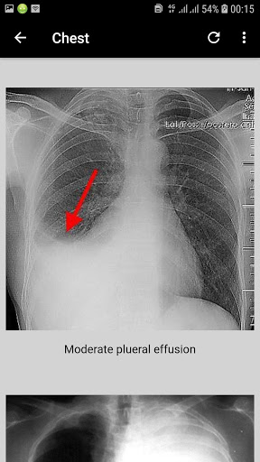
CHEST X-RAY (CXR) INTERPRETATION
Chest x-rays (CXR) are a frequently performed radiological investigation that you’ll be expected to be capable of interpreting (as due to the sheer volume of chest x-rays requested they are often not reported immediately). Therefore before hitting the wards as doctors, it is essential that you develop the ability to interpret chest x-rays, of particular importance, is the ability to recognise findings that require immediate medical attention.
Confirm the following details - x-ray interpretation guide :
Always begin by checking the following:
Patient details (name / DOB)
Date and time the film was taken
Any previous imaging (useful for comparison)
Assess image quality :
Then briefly assess the quality of the image: A mnemonic you may find useful is ‘RIPE’:
Rotation :
The medial aspect of each clavicle should be equidistant from the spinous processes
The spinous processes should also be in vertically orientated against the vertebral bodies.
Inspiration
5-6 anterior ribs, the lung apices, both costophrenic angles and lateral rib edges should be visible
Projection
AP vs PA film :
Tip- if there is no label, then assume it’s a PA. Also, if the scapulae are not projected within the chest, it’s PA.
Exposure :
Left hemidiaphragm visible to the spine and vertebrae visible behind heart
CXR interpretation (ABCDE approach) :
Airway
Trachea
Is the trachea significantly deviated?
The trachea is normally located centrally or just slightly off to the right
If the trachea is deviated, look for anything that could be pushing or pulling at the trachea.
Also inspect for any paratracheal masses/lymphadenopathy
Pushing of trachea – e.g. large pleural effusion / tension pneumothorax
Pulling of trachea – e.g. consolidation with lobar collapse
Rotation of the patient can give the appearance of a deviated trachea, so as mentioned above, check the clavicles to rule out rotation as the cause
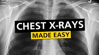
How to Read a Chest X-ray – A Step By Step Approach
There is no perfect way to read an x-ray. However, the important message I would like to give is, to adopt one or the other approach, and to use the chosen approach consistently.
On all Xrays check the following:
Check patient details
First name, surname, date of birth.
Check orientation, position and side description
Left, right, erect, ap, pa, supine, prone
Check additional information
inspiration, expiration
Check for rotation
measure the distance from the medial end of each clavicle to the spinous process of the vertebra at the same level, which should be equal
Check adequacy of inspiration
Nine pairs of ribs should be seen posteriorly in order to consider a chest x-ray adequate in terms of inspiration
Check penetration
one should barely see the thoracic vertebrae behind the heart
Check exposure
One needs to be able to identify both costophrenic angles and lung apices
9Apps 4.9